
Abdomen / Pelvis
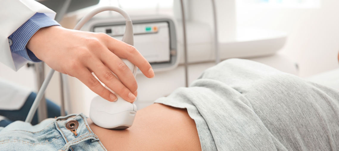
Abdominal ultrasound imaging is performed to evaluate organs such as the kidneys, liver, gallbladder, pancreas and spleen. It can help to diagnose a number of conditions including abdominal pains, inflamed appendix, enlarged abdominal organ or aneurysm in the abdominal aorta or stones in the gallbladder or kidney.
For this type of scan you will need to tell the sonographer if you have had a barium enema or a series of upper GI (gastrointestinal) tests within the past two days as barium remaining in the intestines can interfere with the ultrasound test.
For a study of the liver, gallbladder, spleen, and pancreas, you may be asked to eat a fat-free meal on the evening before the test and then to avoid eating for eight to 12 hours before the test.
For ultrasound of the kidneys, you may be asked to drink four to six glasses of liquid about an hour before the test to fill your bladder. You may be asked to avoid eating for eight to 12 hours before the test to avoid gas buildup in the intestines.
For ultrasound of the aorta, you may need to avoid eating for eight to 12 hours before the test.
You will lie on your back on an examination couch and the transducer moved back and forth across your abdomen to image the area of interest. You may be asked to turn on your side or sit up. You may also be asked to take a deep breath in and hold it for a short while in order to obtain the best possible images
Musculoskeletal
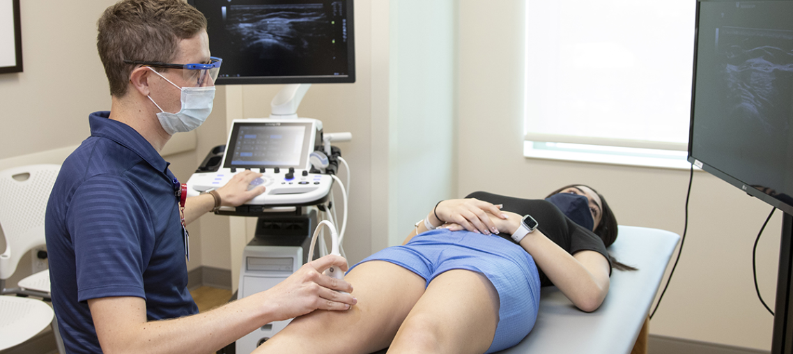
Ultrasound images of the musculoskeletal system provide pictures of muscles, tendons, ligaments, joints and soft tissue throughout the body. This type of scanning is typically used to assist in the diagnosis of tendon tears, such as tears of the rotator cuff in the shoulder or Achilles tendon in the ankle, abnormalities of the muscles, such as tears and soft-tissue masses, bleeding or other fluid collections within the muscles or damage to the major joints. It can also be used as a guide for steroid injections into joints and surrounding soft tissues as an effective treatment for painful joints. No special preparation is required for these types of scan.
At the examination you will need to remove all clothing and jewelry in the area to be examined and may be asked to wear a gown during the procedure. Usually you will lie on your back on an examination couch, but for specialised areas you may be in different positions. The transducer will be moved back and forth across the relevant part of your body on order to image the area of interest. You may also be asked to move joints or limbs during the scanning so that the sonographer can look at the affected area while it is in motion.
Obstetrical
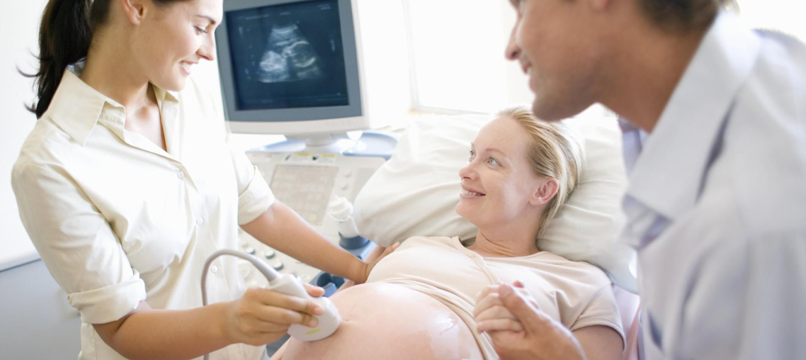
For all Obstetrics scans, no special preparation is needed, although it is best to wear loose clothing that can easily be lifted or removed in order to expose your abdomen. You will lie on your back on an examination couch and the transducer moved back and forth across your stomach in order to gain the best possible image of the fetus.
Ultrasound imaging in pregnancy is widely used to evaluate the baby. It can determine if a baby is present, the position of the fetus and if there is a multiple pregnancy. It can also help to diagnose abnormalities or problems, help determine the age of the pregnancy and subsequent due date as well as showing the position of the placenta in relation to the birth canal.
The Royal College of Obstetricians & Gynaecologists (in their 2000 document on Ultrasound Screening), recommend an Early Pregnancy Scan, undertaken before 15 weeks, which can combine the functions of the Early Viability Scan, Nuchal Translucency Scan and dating scan. There is also then a routine scan at 20 weeks. These two scans are the ‘minimum’ number of scans required during pregnancy and are offered by ¾ of ultrasound units in the UK. Individual circumstances may dictate that more scans may be offered and a breakdown of what you could receive is detailed below.
Small Parts
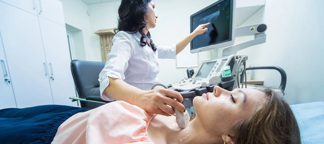
Ultrasound imaging of small parts is a useful way of evaluating many of the superficial organs of the body such as the thyroid gland, the neck and the testes. The ultrasound images can help an ultrasound practitioner to evaluate these organs and look for signs of disease.
Abdomen / Pelvis
Abdominal ultrasound imaging is performed to evaluate organs such as the kidneys, liver, gallbladder, pancreas and spleen. It can help to diagnose a number of conditions including abdominal pains, inflamed appendix, enlarged abdominal organ or aneurysm in the abdominal aorta or stones in the gallbladder or kidney.
For this type of scan you will need to tell the sonographer if you have had a barium enema or a series of upper GI (gastrointestinal) tests within the past two days as barium remaining in the intestines can interfere with the ultrasound test.
For a study of the liver, gallbladder, spleen, and pancreas, you may be asked to eat a fat-free meal on the evening before the test and then to avoid eating for eight to 12 hours before the test.
For ultrasound of the kidneys, you may be asked to drink four to six glasses of liquid about an hour before the test to fill your bladder. You may be asked to avoid eating for eight to 12 hours before the test to avoid gas buildup in the intestines.
For ultrasound of the aorta, you may need to avoid eating for eight to 12 hours before the test.
You will lie on your back on an examination couch and the transducer moved back and forth across your abdomen to image the area of interest. You may be asked to turn on your side or sit up. You may also be asked to take a deep breath in and hold it for a short while in order to obtain the best possible images